Identify The Structures Labeled In The Diagram. Label A Solv
Solved drag the labels onto the diagram to identify the Acids proteins biology carboxyl gabi expii Human epidermis diagram human skin diagram human skin photo & picture
Solved 104 Review Sheet 7 4. Lobal the bi Label the skin | Chegg.com
Parts of a plant cell labeled Solved identify the labeled structures in the diagram below chegg com Drag the labels onto the diagram to identify the structures and
Macromolecule acid amino protein structure chemical chemistry macromolecules dna general basic major alpha chapter group labeled clipart acids building figure
Part a label the appropriate structures on thisLabeled prokaryotic cell diagram Drag the labels onto the diagram to identify the structural elements ofDrag onto transcribed.
Art labeling activity the structure of a skeletal muscle fiber diagramLabel the parts of a neuromuscular junction Solved label the appropriate structures on this diagram withSolved drag the correct labels onto the diagram to identify.

Amino acids acid group carbon chain side central carboxyl variable atom hydrogen asymmetric generic molecules lecture aminoacid libretexts which reading
Chapter cells drag labels appropriate targets respective their living units printSolved art-labeling activity: the anatomy of the ear Solved 104 review sheet 7 4. lobal the bi label the skinA&p lab figure 8.3 structure of a long bone pt 1 diagram.
2017_ss1_lecture_03Neural stimulation of muscle contraction Solved dna replication drag the labels to their appropriateSolved drag the labels onto the diagram to identify the.

Solved drag the labels onto the diagram to identify the
Labeling drag ontoSkin label section vertical layer subcutaneous using provided terms chegg system answers dermis question integumentary sebaceous glands biology sweat transcribed Intestine small parts diagram three anatomy system digestive humanSkin structure diagram epidermis anatomy human label face histology physiology foot function care picture choose board stratum corneum facial separate.
Muscle contraction reticulum sarcoplasmic skeletal diagram stimulation neural steps acetylcholine action potential cell muscles calcium synaptic excitation cross figure membraneCh103 – chapter 8: the major macromolecules – chemistry Solved label the vertical section of the skin andSolved art-labeling activity: structure of the epidermis.

Three parts of small intestine diagram
Review sheet art-labeling activity 52 of 4 a drag the labels onto theProteins — overview & importance in biology [solved] drag the labels onto the diagram to identify the structuresLabeling epidermis targets respective appropriate homework.
Solved drag the labels onto the diagram to identify theAnswered: label the figure to assess your… Sheet skin label review structures diagram indicated areas integumentary answers accompanying solved lobal bi systemPrint a&p chapter 3 cells: the living units flashcards.

Diagrams: heart nerve control dirgram
External ear diagram labeledIntegumentary system labeling worksheet The diagram below shows a bacterial replication fork and[solved] art-labeling activity: figure 13.13a drag the appropriate.
.
![[Solved] Art-labeling Activity: Figure 13.13a Drag the appropriate](https://i2.wp.com/www.coursehero.com/qa/attachment/37180819/)

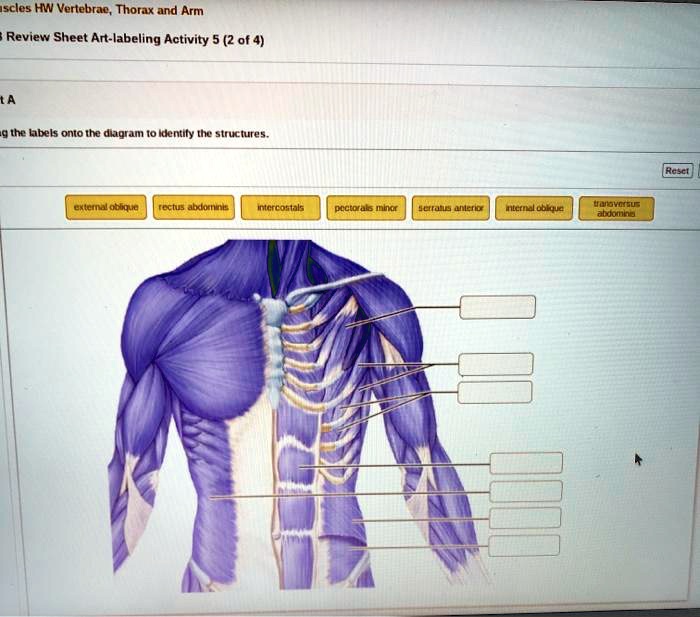
Solved Drag the labels onto the diagram to Identify the | Chegg.com

Art Labeling Activity The Structure Of A Skeletal Muscle Fiber Diagram

Human Epidermis Diagram Human skin diagram human skin Photo & Picture

Answered: Label the figure to assess your… | bartleby
Review Sheet Art-labeling Activity 52 of 4 A Drag the labels onto the

Solved Drag the labels onto the diagram to identify the | Chegg.com

Three parts of small intestine diagram